Illuminating a ubiquitin transfer relay, step-by-step
Commentary on Simona Polo's paper published on Nature Structural & Molecular Biology
April 2014
A key way that protein activities are controlled in a spatiotemporal manner is by post-translational modification, such as by covalent ligation of the small protein ubiquitin. In some cases, a single monoubiquitin is attached. Alternatively, repeated rounds of ubiquitin ligation can form polyubiquitin chains with multiple ubiquitin molecules attached to each other. Chains can form on several different sites of ubiquitin.
Homologous to E6-AP C terminus (HECT) E3 ligases recognize and directly catalyze ligation of ubiquitin (Ub) to their substrates. Molecular details of this process remain unknown. We report the first structure, to our knowledge, of a Ub-loaded E3, the human neural precursor cell-expressed developmentally downregulated protein 4 (Nedd4). The HECT(Nedd4)~Ub transitory intermediate provides a structural basis for the proposed sequential addition mechanism. The donor Ub, transferred from the E2, is bound to the Nedd4 C lobe with its C-terminal tail locked in an extended conformation, primed for catalysis. We provide evidence that the Nedd4-family members are Lys63-specific enzymes whose catalysis is mediated by an essential C-terminal acidic residue.
[PMID: 23644597]
Initially, polyubiquitin chains were mainly known for mediating interactions with the proteasome and directing degradation of the modified protein. However, it is now known that chains linked via lysine 63 on ubiquitin ("K63-linked chains") regulate diverse functions such as protein trafficking, DNA repair, and immune signaling. Likewise, monoubiquitin can bestow its targets with a wide-range of alternative fates. Therefore, a major question for understanding protein regulation is: how are specific ubiquitin modifications directed to their particular protein substrates? Simona Polo at IFOM and I came to work on this question from different directions. I have long been interested in the biochemical and structural basis by which ubiquitin becomes attached to targets to regulate function. For over a dozen years, Simona Polo has been a leader in understanding numerous facets of ubiquitin signaling, although her path started with a quest to decipher regulation of and by the epidermal growth receptor. In seminal work performed initially with Pier Paolo Di Fiore, and then later in her own lab, Simona found that Kay Hofmann's predicted ubiquitin interacting motif (UIM) sequence in epsin family endocytic regulators both binds monoubiquitin, and is also responsible for Eps15 monoubiquitination. Simona's lab discovered a novel mechanism termed "coupled monoubiquitination", where Eps15 uses its UIM to bind ubiquitin on an E3 enzyme NEDD4. The burning follow-up problem was to understand how NEDD4 transfers ubiquitin.
NEDD4 belongs to the family of E3 enzymes known as HECT. NEDD4 and other HECT E3s function as part of multi-enzyme ubiquitin transfer cascades through a two-step mechanism. First, ubiquitin is transferred from the catalytic cysteine of an E2 enzyme to that on the E3 HECT domain. This produces a transient HECT~ubiquitin intermediate that is linked by a thioester bond. Next, ubiquitin is transferred from the HECT E3 cysteine to either a substrate protein, or to a specific lysine on the surface of ubiquitin to generate a chain. This process is often likened to a relay, where ubiquitin is the baton transferred between athletes (e.g. E2, E3, substrate, and eventually ubiquitin linked to substrate). We had previously determined the structure transfer of ubiquitin from E2 to the HECT domain and subsequently of a downstream intermediate, and Simona's and Jon Huibregtse's labs had solved structures of other forms of HECT E3s bound to ubiquitin.
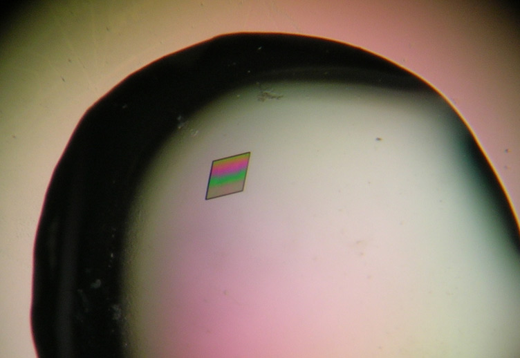
However, several big questions remained. In particular, noone had ever been able to "see" the transient intermediate with ubiquitin linked at a HECT E3 catalytic cysteine, because such complexes are extremely unstable. Simona and her colleagues overcame this challenge with a very clever approach, by using an alternative, more stable bond to link the active site of the NEDD4 HECT E3 and ubiquitin. Their study published in Nature Structural and Molecular Biology was a big breakthrough, and remains to date the only structure capturing an elusive HECT E3~ubiquitin intermediate, as if after ubiquitin transfer from E2! Their data beautifully shows a mechanism for how the E2-to-E3 relay goes forward even though the thioester bonds linking E2~ubiquitin and HECT E3~ubiquitin intermediates may be perceived as similar. In the context of the HECT E3~ubiquitin intermediate, there is a new beta sheet formed between NEDD4 and ubiquitin, which stabilizes the complex. In terms of visualizing ubiquitin transfer cascades, our combined data suggest that NEDD4 functions as if in a children's relay. First NEDD4 faces E2 to receive ubiquitin. After ubiquitin transfer to the NEDD4 HECT domain, a new beta sheet stabilizes the intermediate. Then, the NEDD4 HECT E3 turns around to face the substrate.
The article from Simona's group also reports that all HECT E3s related to NEDD4 build K63-linked chains. These may mediate trafficking through membrane compartments, or regulate substrates through other proteasome-independent means. The authors also found residues that when mutated completely eliminate K63 chain formation. This will serve as a very useful tool for numerous researchers trying to unravel myriad functions of HECT E3s in cells. It also serves as tremendous motivation to understand how and why NEDD4 builds K63-linked ubiquitin chains.
On a personal note, Simona's enthusiasm is infectious, and it has been immensely inspiring for me to share data and discuss ideas with her along my own path.


